
No doubt you've heard of these and other fun midwives' tales of gender prediction. They are all exactly 50% accurate (which is the same accuracy as your own guess!).
Is there a way to know your baby's gender before the mid-pregnancy ultrasound (around 18-20 weeks)?
You may actually have a chance...
The risky way:
CVS (Chorionic Villus Sampling) and Amniocentesis are two tests that occur before 20 weeks, and your doctor may recommend them if there is reason to suspect any defect or disorder in the baby. They are invasive tests that will allow specialists to examine the baby's DNA. Of course, that means they can also see whether the baby is a boy or a girl. NOTE: Knowing the gender early is not a good enough reason to have these tests performed. They both carry some serious risks.
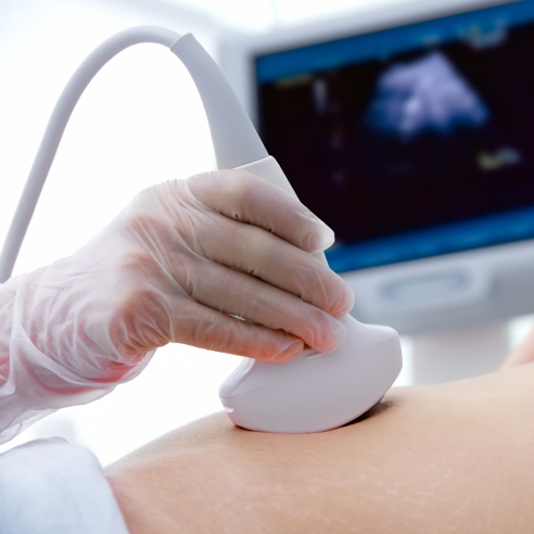
The safer way:
A much safer option is what is known as the Ramzi method. Dr. Saad Ramzi Ismail, an ultrasound supervisor and instructor, finished a study in 2007 that concluded:
At 6 weeks gestation, the gender of a baby can be determined with 97% accuracy by the location of the chorionic villi (the forming placenta) in the uterus. This study was done with ultrasounds, watching for special markers that are used for by ultrasound techs. (If you want to know more specifics so you can ask your ultrasound tech about it, here is a good summary of the study, or if you want even more detail, you can see a copy of the study at the bottom of this post, borrowed from OBGYN.net).
In a very small nutshell, the study concluded:
Placenta on the right side of the uterus:
BOY (97.2% accurate)
Placenta on the left side of the uterus:
GIRL (97.5% accurate)
Keep in mind, that doesn't mean what side your baby is on when they look at the ultrasound. It means on which side of the uterus the placenta is beginning to form. It may be impossible for you to tell, but if your ultrasound tech knows what markers to look for, they should be able to tell you easily. If they have doubts, show them the article!
This study was done at 6 weeks gestation, then checked for accuracy at 20 weeks (97% accurate!). It's a pretty big deal to have such a high accuracy rate.
Leave a comment below: was this study correct in your case?
Helpful links:
What to Expect: Finding out Gender Before 10 Weeks
Summary of Dr. Ramzi's 10 year study
(Here's that study I mentioned. A good read if you can understand the technical terms)
The Relationship Between Placental Location and Fetal Gender (Ramzi’s Method)
By
ORIGINAL RESEARCH
By
Dr. SAAD RAMZI ISMAIL
Ultrasound Supervisor / Instructor
NWHC-High Level Hospital-Alberta
Pobox-1462, High Level, Alberta, T0H1Z0
Canada
DECEMBER 2007
ORIGINAL RESEARCH
By
Dr. SAAD RAMZI ISMAIL
Ultrasound Supervisor / Instructor
NWHC-High Level Hospital-Alberta
Pobox-1462, High Level, Alberta, T0H1Z0
Canada
DECEMBER 2007
Conflict of Interest DeclarationThe author acknowledges no commercial affiliation or financial conflict of interest.
AcknowledgmentThe author wishes to express his appreciation to the project supervisors Dr. Ali Cadili and Dr. Lincoln M Abney PhD. Special thanks to Mrs. Merle McCann, Dr Essam Shaaban, Dr. V Bouta, Dr. S. Desilva, Dr. P. Hughes, Dr. A zammit, and Siti Arabiah Hamid for their help and guidance.
DedicationThis project is dedicated to all those who strive for knowledge and wisdom, to those who believe in the creator and intelligent design, and to the memory of my beloved parents and brothers who were killed in Iraq.
Advances in KnowledgeThis study contributes to advances in knowledge by understanding the history of fetal gender and the ethical dilemma of choosing or detecting fetal gender at first trimester of the pregnancy. It gives new prospective and method to detect fetal gender as early as possible to better manage some genetic disease which can be found in male or female fetuses. Thus, gives the parent the choice of what to do, and gives the gynaecologist’s and genetic counsellors the ability to manage, discuss and guide the parents to better management of the fetus.
This study might be used in the veterinary medicine to help endanger species and to increase the probability of conceiving male or female of certain specious. It also increases the skills of the sonographers and Radiologists to detect fetal gender by applying this method. It might also be used as a genetic soft marker when bilateral pyelectasis is present. This study will opens the door toward new and radical understanding of the female uterus.
Application to Patient CareThe application to patient care is in the knowledge gained by physicians, genetic counsellors and researchers that can be applied from the sonographers/sonoliogists ability to detect fetal gender as early as possible especially in families with genetic disorders that can be found in male or female fetuses. It will enhance the sonographer’s ability to correlate the finding with other genetic soft markers such as renal pyelectasis.
Application to Patient CareThe application to patient care is in the knowledge gained by physicians, genetic counsellors and researchers that can be applied from the sonographers/sonoliogists ability to detect fetal gender as early as possible especially in families with genetic disorders that can be found in male or female fetuses. It will enhance the sonographer’s ability to correlate the finding with other genetic soft markers such as renal pyelectasis.
This study may help parents to decide and choose the type of medical management available in case of inherited genetic problem such as in X-linked genetic disorder.
This study might not be for all patients but definitely for those who want to know and prepare their life and finances and for those who have a genetic problem, which can inflect one gender than the other.
This study might not be for all patients but definitely for those who want to know and prepare their life and finances and for those who have a genetic problem, which can inflect one gender than the other.
Abstract The aim of this study is to determine the relationship between placental /chorionic villi laterality and fetal genders early in pregnancy using 2-D ultrasonography and color flow Doppler.
Material and MethodThis is a multi-center prospective cohort study of 5376 pregnant women that underwent ultrasonography from 1997 to 2007. Trans-vaginal sonograms were performed in 22% of the patients at 6 weeks gestation, and Trans-abdominal sonograms were used at 18-20 weeks gestation, at this time the fetal gender were confirmed in 98-99%. The fetal sex was confirmed 100% after delivery. The study also addressed the bicornuate uteri with single pregnancy in relation to placenta / chorionic villi location. The result was tabulated according to gender and placenta / chorionic villi location. Bicornuate uteri with single fetus in different horns were studied and tabulated
Result Dramatic differences were detected in chorionic villi / placental location according to gender. 97.2% of the male fetuses had a chorionic villi/placenta location on the right side of the uterus whereas, 2.4% had a chorionic villi/placenta location to the left of the uterus. On the other hand 97.5% of female fetuses had a chorionic villi/placenta location to the left of the uterus whereas, 2.7% had their chorionic villi/placenta location to the right side of the uterus.127 cases were found to involve bicornuate uteri with single foetuses, most male fetuses were located in the right horn of the uterus and showed right placental laterality (70%). Most female fetuses 59% on the other hand, were located in the left horn and showed left laterality (59%).Moreover, most of the males located in the left horn exhibited right laterality (89%). Also most females located in right horn exhibited left laterality (976.4%). In addition this research indicated that there was a possible link between renal pyelectasis and placental location, and it might be used as a genetic soft marker.
ConclusionRamzi’s method is using placenta /chorionic villi location as a marker for fetal gender detection at 6 weeks gestation was found to be highly reliable. This method correctly predicts the fetus gender in 97.2% of males and 97.5% of females early in the first trimester. And it might be helpful to use as a genetic soft marker in relation with fetal pyelectasis.
INTRODUCTION
There are no literatures regarding the placental or chorionic villi site in relation to fetal gender.
Adequate visualization of the fetal gender is feasible by high-resolution real-time ultrasonography during the prenatal examination (Figure 1, 6, & 7). Technical difficulties that were reported relate to fetal presentation, number of fetuses, fetal activity, amniotic fluid volume, and maternal obesity or bowel gas. In the current study, the fetal position, particularly breech presentation and oligohydramnious were the major factors obscuring sex determination1-8. Although fetal sex can be determined as early as 13 to 14 weeks, most sonographers agree that the sonographic detection rate sharply increases after 18 weeks of gestation. Some reasons of detecting fetal gender are abnormal genitalia in X-linked disorders, testicular feminization, pseudohermaphroditism, hydrocele and more (Figure 2, 3) 1_8. Fetal sex can be detected sonographically and the genitalia can be predicted successfully 83.5% of the time between 16-20 weeks gestation9.
Determination of fetal sex is not only done for parental curiosity but also has many medical advantages. Accurately assessing fetal sex can assist in assigning zygosity in twin pregnancies. Ambiguity of the genitalia can occasionally be detected sonographically after detecting other abnormalities (Figures 8, 9), because of a relevant family history. Some cases are diagnosed after careful evaluation of fetal gender because of an antenatal discrepancy between the fetal karyotype and the genital anatomy. In women at risk of X-linked genetic disease or ambiguous development of the external genitalia early gender assignment may give parents the option to avoid invasive testing in up to half of cases where the fetus would not be affected 1_8. Sonographers might confuse the Vestigial tail in a female fetus with a male fetus (Figure 11), or the 3 lines sign of the male skin folds with a female fetus (Figure 23).
Human gonads of both sexes begin developing at 4 weeks of gestation. At this early stage they are identical and capable of differentiating into either testes or ovaries 10. They are situated next to the mesonephric or Wolffian duct and the paramesonephric or Mullerian duct, which are prominent in the development. The wolffian duct developed into the male and female reproductive system respectively 11.
Testicular differentiation begins at » 7 weeks and is regulated by the testes-determining factor, a gene located on the short arm of the Y chromosome. Production of anti-Mullerian factor by Sertoli cells in fetal testes inhibits Mullerian duct development of the Wolffian duct into epididymis, vas deferens, and seminal vesicles. In fetuses with a normal ovary, or absence of any gonad, the Mullerian duct develops into Fallopian tubes, uterus, and upper vagina 10, 11.
Up to 11 weeks of gestation the growth and the development of the external genitalia is identical in both sexes. After this there is rapid differentiation of the genital tubercle into the male or the female phallus. As significant differences in the rate of penile and clitoral growth only become evident after 14 weeks, when most of the prenatal growth of the penis occurs, evaluating phallic size before this time will lead to erroneous gender assignment (Figures 4, 5).
Examined in the transverse section the clitoris appears similar to the penis in early pregnancy, but in the mid-sagital sections the direction in which the genital tubercle points can be evaluated and this differs in males and females 12, 13.
Scientists indicated that in the male fetus the orientation of the phallus is anterior, perpendicular to the lumbosacral spine and in the female fetus the orientation is caudal, parallel to the axis of the lumbosacral spine. The orientation of the phallus in early pregnancy was termed the sagital sign and several authors have used various permutations of this sign to attempt accurate gender assignment in early pregnancy (Figures 12, 13, 14, &15) 14_18.
Hypospadias detected by prenatal US is usually of the severe type, although occasionally less severe forms can be detected when the external genitalia are carefully examined after detecting associated urogenital tract anomalies 19. Meizner (2002) described the “tulip sign” as an ultra-sonogram clue for the in-utero diagnosis of severe hypospadias, the “tulip: being formed by the ventrally orientated penis located between the scrotal folds (Figure 10) 19.
The use of the phallus angle as prediction for fetal sex was studied by Efrat (1999) in 172 women using a midline sagital scan with the fetus supine and the spine parallel to the transducer face (sound lines perpendicular) ‘without extension of the spine or limbs’, angles were drawn on an image of the genital area with a line through the genital tubercle and another through the lumbosacral skin line, his study concluded that “a final decision on invasive testing for sex-linked conditions should be undertaken only after 12 weeks of gestation (Figure 12,13,14,&15) 15.
Ultrasound prediction of fetal gender study was conducted at 14-20 weeks gestation in 843 fetuses; it showed an error of 93.3% in diagnosis of gender were more likely to occur in the assessment of the female fetus 20.
Sex chromosomes can be affected through changes in the environment in which the genes are expressed. Diet and food with certain minerals play a big role in this theory 21.
Some abnormalities of the phallus could be related to other genitourinary tract malformations, such as dilatation of upper urinary tract, congenital megalourethra is sometimes associated with prune-belly syndrome 22.
The diagnosis of hypospadias possibly lead to diagnosis of other pathology such as cleft lip and palate, and opitz’s diseases (hypertelorisim and hypospadias), which is inherited as an autosomal dominant disorder 23. In addition a study by Jones (1988), suggested that other autosomal recessive disorder in which hypospadias is known to occur is Smith-Lemli-Optiz syndrome, which consists of syndactyly of the toes, microcephaly, moderately severe mental deficiency and failure to thrive 24.
Determination of fetal sex at first trimester is very important. In case of pregnancies at risk for congenital adrenal hyperplasia, to stop the dexamethasone(Drug information on dexamethasone) treatment to female fetuses resulting in a substantial decrease of unnecessary treatment and invasive diagnosis tests 25.
According to coral reef studies report (2000) in Australia, Coral fish, Clown fish, hard clam, and goper fish, they all started as male. They can switch to female later when the fish get older. It switches to female to keep producing eggs. When they change their sex, they cannot go back. Some Clown fish can switch back and forth female to male or opposite, while turtle and alligators can either the sex of their hatching by changing the temperature and the humidity as they can put their eggs deeper or shallower in the sand farther or closer from the water 26. A male fetus with a micro penis and undescended testicles cannot be distinguished from a female who has clitoral hypertrophy 27.
The hardest diagnosis even for an experienced sonographer is visualization of ambiguous genitalia. Ambiguous genitalia are defined as according to Mosby’s (1994) “External genitalia that are not normal and morphologically typical of either sex.” Ambiguous genitalia can be the result of many underlying conditions 28.
Embryology and the Determination of Sex
The normal amount of chromosomes in humans is 44. With the addition of two sex chromosomes and the normal male has one X and Y chromosome. Abnormalities can occur during any stage of meiosis (when sex cells divide) or in the first mitotic division in the zygote, which could result in having more or less in numbers of sex chromosomes. Any abnormalities in the sex chromosomes number are associated with some abnormality in the developing cells 29.
The development of gonads is a unique embryological situation. Any other type of embryological developing organ can only develop into one type of organ (e.g. heart, spleen, stomach). The developing gonads have two options; they can develop into either an ovary or testis. Before the gonads can develop into either, the gonad goes through an indifferent stage-when it is neither female nor male 30, 31
Both males and females initially have the same two pairs of ducts the mesonephric ducts (Wolffian) and the paramesonephric ducts (Mullerian). One pair degenerates while the other pair develops into male or female anatomy 31. If the fetus is going to be male, the sex cords continue to proliferate and form a medullary, which become the rete testis. The Wolffian duct develops into the vas deferens and the Mullerian duct degenerates. In females, the Mullerian duct develops and the Wolffian duct degenerates. The Mullerian duct at about 9 to 12 weeks gestational age will fuse caudally and develop into the uterus, fallopian tubes and 2/3 of the upper vagina31.
Abnormalities
A malfunctioning adrenal gland can cause a genetically female baby to be exposed to levels of the male hormone testosterone, which would be more typically found in a baby boy. The result may be a baby girl with an unusually large, even penis-like clitoris and sometimes with labia that look like a scrotum 29, 30.
According to Potter (1975), there are number of syndromes in which there is a deviation from the normal internal and external genitalia and these are:
Turner’s syndromeTurner’s syndrome is an extremely common chromosomal anomaly occurring in about 1 in 3000 live female births.
Klinefelter’s SyndromeKlinefelter’s syndrome is syndromes of gonadal defects appearing in males who have an extra X chromosome, making there karyotype typically XXY. Characteristically these infants have small firm body and long legs.
Testicular Feminization SyndromeTesticular feminization syndrome occurs in genetic males with morphologically recognizable testes.
Female Pseudo-hermaphroditesAccording to Mosby (1994), the ones affected by female pseudohermaphroditism are genetic females (46XX) with congenital adrenal hyperplasia and abnormal androgen production 28.
Male PseudohermaphroditismMale pseudohermaphroditism is a rare genetic defect that leads to feminization of the genitalia28.
True HermaphroditesHermaphroditism is a very rare condition in which both testicular and ovarian tissue exists in the same person28. Other minor reasons to know the gender is that a splenia (absence of spleen) is more common in males while Polysplenia (multiple spleens) syndrome is more predominant in females32, 33.
True HermaphroditesHermaphroditism is a very rare condition in which both testicular and ovarian tissue exists in the same person28. Other minor reasons to know the gender is that a splenia (absence of spleen) is more common in males while Polysplenia (multiple spleens) syndrome is more predominant in females32, 33.
The Placenta Formation
The amnion is a membranous sac that surrounds the embryo (later on the fetus). Early in the development of fertilized ovum, when it forms a ball like structure, in the blastula, as slit like cavity appears between the embryonic disc (the embryo proper) and the trophobliastic covering which invade the uterine wall. By the seventh day, a roof appears which is made of thin sheet of cells probably derived from the cytotrophoblasts. The floor of the cavity is made up from the ectoderm (outer layer) of the embryonic disc. As the amnion enlarges, it gradually obliterates the chorionic cavity (which forms the second sac), and sheaths the umbilical cord. The sac becomes filled with a watery fluid (98% water) that is derived from the maternal blood by transport across the amnion 6, 34, 35.The chorionic villi are the functional endocrine units of the placenta. An inner layer, cytotrophoblasts, and an outer layer, syncytiotrophoblast surround a central core with abundant capillaries that makes it helpful to detect using color flow and angio power Doppler (Figures 24, 25). The inner layer produces neuropeptides, while the outer layer produces the protein hormones; human chorionic gonadotropin (HCG) and human placental lactogen (hPL); and the sex steroids: estrogens(Drug information on estrogens) and progesterone(Drug information on progesterone) 35, 36, 37.
According to Kavraiska N (1993) the temperature does increase in the placenta compared to the uterus due to change in the placental blood flow hemodynamic 38. And that the right side of the uterus have more blood flow than the left side 38.
According to Kavraiska N (1993) the temperature does increase in the placenta compared to the uterus due to change in the placental blood flow hemodynamic 38. And that the right side of the uterus have more blood flow than the left side 38.
CHORIONIC VILLI SAMPLING (CVS)
Chorionic villi sampling is an invasive procedure can detect the fetal gender as early as 11 weeks. Chorionic villus sampling (CVS) has come to be regarded as a safe and effective diagnostic procedure for the diagnosis of fetal chromosome abnormalities and single gene disorders 39, 40.
GENDER AND THE FUTURE
“What’s next in sex selection and sex detections? Will there be a 100 percent effective method soon?” Sex selection in the near future, say the next ten years, will probably be much as it is now. Better methods of separating the two types of sperm may enable doctors to improve on current techniques somewhat-but only with methods that use artificial insemination. Researchers have recently reported sperm-separation techniques that provide concentrates of female-producing sperm, though these remain experimental. Scientists use a separation technique that exploits the tiny differences in the surface electrical charge of the two types of sperm 41, 42.
Looking further ahead into the future, Dr. E. J. Leiberman, formerly of the National Institutes of Health, once suggested that women might eventually have “a special diaphragm that will let through only the sperm that carries, let’s say the male sex and hold back those that carry the female sex.” Dr. Shettles did, in fact report some years ago, in the Journal of Urology, that he had been able to devise filters that would permit passage of the male-producing sperm while restraining most of the female sperm.
Dr. Charles Birch, while head of Sydney, Australia University, School of Biological Science, predicted that researchers would eventually come up with chemical packaged in the form of pills that would determine sex. If male offspring were desired, Dr. Birch said, the husband would simply take one of the “little boy pills” just before intercourse or if a daughter was wanted, one of the “little girl pills. In 1995, researchers at John Hopkins, Duke University, and elsewhere reported that sperm have olfactory receptors that enable them to “sniff” or “smell” the egg – and that they may be drawn toward it by “chemo attraction.” This new finding excited a lot of attention on the media.
In late 1987, researchers affiliated with the Massachusetts Institute of Technology reported that they believe they have found the “trigger” – a single gene –within the Y chromosome that determines male sex; it is the absence of this gene in the X chromosome that results in development of female offspring.
Uterine Contraction
Uterine contraction begins asymmetrically a “pacemaker” in one uterine horn, and that progesterone from the placenta blocks myometrium contractibility primarily at the site of implantation. Case records were examined at 182 patients who had placental locations performed and who had a spontaneous onset of labour Where the placenta was implanted in the right upper quadrant of the uterus labour occurred on average four days sooner than when it was implanted in the left upper quadrant” the difference was statistically significant 43.
This theory also mentioned by Larks work on the human electrohysterogram. Larks proved that electricity create contraction or expansion of the uterine muscles on his study of 293 subjects in labour using ECG –bipolar electrodes 44.
Monitoring of uterine contraction activity is an important diagnostic tool used during both pregnancy and labour. The strain the pregnant uterus exerts on the maternal abdomen is measured via external tocography 45. However, limitation of this approach has caused the development of another technique-electohysterography - that is based on the recording of electrical uterine activity. The comparison study between electrohysterography and tocography was carried out thanks to the possibility of simultaneous recording of mechanical and electrical uterine activities 44, 45.
Endometrial wave like movements have been characterized and found to be of relevance to fecund ability during the natural ovarian cycle 46, 47, 70. Researchers found evidence that a greater frequency of contractions on the day of embryo transfer appears to be associated with a reduced pregnant rate but others observed the converse46, 47, 48. To describe endometrial contractility, the term junctional zone contraction is used because there is evidence that this particular layer of myometrium, which consists of a discrete compartment of more compacted myocytes, may be responsible for the wavelike movements of the adjacent endometrial 49, 50.
Polarity and Cellular Communication
The polarity cycle which unknown to most of us was there in addition to the ovulation/menstrual cycle .When the ovum membrane is neutral; sperms carrying either the X or Y chromosome can unite with the ovum and fertilize it51. During this period, the chances of getting a baby boy or baby girl are 50: 50. When the ovum, membrane is positively charged, it attracts the sperm carrying X chromosome (which is negatively charged) and a baby girl is produced. Scientist researching cellular communications has found that the mother’s ovum membrane’s receptors had an alternating polarity (energy) that accepts or rejects the “x” or “y” chromosome sperm according to her energy cycle 51, 52.
Researchers found that the “y” chromosome sperm had a constant different charge to “x” chromosome sperm 51, 52. French scientists went further and verified that the ovum membrane receptors produce a different and alternating polarity that occurred as a result of a mother’s biological clock that they call the polarity cycle 51, 52, 53. The ovum membrane’s alternating polarity is nature’s way of selecting sperm with different gender determining chromosomes52, 53.
Genes, Hormones and Sex Determination
Researchers know at present that gender determination in man has a genetic origin, but there are other mechanisms that apply in some animals. It appears, for instance that sex may be determined by the incubation temperature of the eggs, which can modify the action of enzymes such as aromatase. Elsewhere, similar systems may have evolved separately and are found in widely divergent species 54, 55, 56. This is called concerted evolution.
Databases searches seeking similar sequences in humans have found a human cDNA that encodes a protein with a DM domain. Moreover, the expression of this protein is seen only in the testis. It is been called DMT1 (DM-Testis) 54, 55, 56.
Moreover, this gene appears to be critical in human sex determination, since mutations in this region is associated with male to female sex reversal. DMT-1 related sequences have also been found in the chick of alligator, and mouse. The DMT genes are expressed only in the genital ridges of male embryos. This suggests that male sex determination using a common gene in sex determining mechanisms in both vertebrates and invertebrates 54, 55, 56.
Sex hormones influence the biological activities throughout our lives, beginning with signaling for certain fetal tissues to differentiate into structures that are specific for male or female 57.
Sex hormones influence the biological activities throughout our lives, beginning with signaling for certain fetal tissues to differentiate into structures that are specific for male or female 57.
Dr Gellersen (1994), study showed that the regulation and function of a newly discovered mammalian polypeptide hormone, stanniocalcin (STC). SCT was originally described as a potent calcium-lowering hormone in fish and has only recently been discovered in mammals and their studies have shown mammalian STC is most highly expressed in the ovary 58.
Male and Female Sperms
Scientists can now identify the male sperm (i.e. carrying Y chromosome) from female sperm (i.e. carrying X chromosome) by means of fluorescent material that attaches itself to the Y chromosome only under the ultraviolet light; the Y carrying sperms fluoresce and glow. According to Hamilton and Mossman (1992), the sex of aborted fetus at six weeks cannot be determined by histological examination of the gonads 59.
Sex Chromosomes
Sex not always determined by sex chromosomes but sometimes by environmental triggers 60, 61. The nuclei of human cells contain 22 autosomes and 2 sex chromosomes. In females, the sex chromosomes are the 2X chromosomes. Males have one X chromosome and one Y chromosome. The presence of the Y chromosome is decisive for unleashing the developmental program that leads to a baby boy 60, 61, 62. Today the X chromosome is still home to thousands of genes, but the Y chromosome has only a few dozen. Of those, 19 are shared between the X and Y 60, 61, 63, 64. These 19 genes are essentially living fossils in that they are able to provide scientist with information about the sex chromosomes and their evolutionary history as well 60-64.
Implantation
When the embryo reaches the uterus it must hatch from the zona pellucida in order to implant. The zone pellucida keeps the embryo from implanting in the walls of the oviduct. When this happens it is called a tubal pregnancy or an ectopic pregnancy and can be very dangerous for the mother. When free of the zona pellucida, the blastocyst can make direct contact with the lining of the uterus (Figures 24, 25) 65, 66. The next major step in human development is called gastrulation. This is the point at which the cells of the embryo form three tissue layers, the endoderm (inside layer), mesoderm, and ectoderm (outside layer). This is one of the most crucial points in development where a great deal of differentiation occurs 67. Maternal effects are broadly defined as the impact that the mother and her phenotype have on the phenotype of the offspring 61, 68. This is quite distinct from the genetic effects that a mother has on her offspring’s phenotype.
Pyelectasis
Fetal Pyelectasis is a pelvicaliceal dilatation. It can be classified according to the gestational age (Figures 19,20) 71.
Between 15 and 20 weeks: greater or equal to 4mm.
Between 20 and 30 weeks: greater or equal to 5mm.
Between 30 and 40 weeks: greater or equal to 7 mm.
Persistent fetal pyelectasis is defined as greater than 7mm in the third trimester 71.
Between 15 and 20 weeks: greater or equal to 4mm.
Between 20 and 30 weeks: greater or equal to 5mm.
Between 30 and 40 weeks: greater or equal to 7 mm.
Persistent fetal pyelectasis is defined as greater than 7mm in the third trimester 71.
Urinary tract abnormalities can be found in up to 5% of newborns; they account for 25% of congenital defects, and contribute to 4% of the prenatal mortality 72. Pyelectasis is usually bilateral (57%), and it is more common in the left kidney 73. Male fetus exhibit a significantly increased frequency of renal pelvic dilatation compared with female fetus at 3/1 ratio 74.
Diagnosis is made by sonographic measurement of the renal pelvic anteroposterior diameter 75.
Diagnosis is made by sonographic measurement of the renal pelvic anteroposterior diameter 75.
Mild pyelectasis is a common finding, with no significant long term effect (Figure 20). However, there is a small association with aneuploidies, especially trisomy 21. The incidence of Down syndrome is 3.3% when pyelectasis are found, and 25% of fetuses with trisomy 21 have pyelectasis 76. The risk of chromosomal anomalies increases when the pyelectasis diagnosed in older women over 35 years of age or if it is associated with other sonographic soft markers 77.
The Ethical Issue of Sex Determination
The moral issues concern whether it is “right” to be determining the sex of a fetus before it is born, and if that knowledge also determines whether or not the baby lives. Using prenatal diagnosis generates information which society has not yet learned to deal with. The issue of who is more at “fault” is also raised. If a fetus is aborted because of its sex, does the responsibility lie more in the hands of the doctor who provided the sex information, or by the doctor who performs the abortion?
The social issues that are raised by sex determination tests; however, this area does overlap into issues of a moral and political nature. In some cultures, the higher status of males goes beyond the male/female salary inconsistencies. Although the approval rate for sex selection (using technologies for sex determination to abort the fetus) is low in the U.S.A, Canada, and The Western countries. Family size is decreasing and there is still a preference for firstborn sons (Boss, p 110). Boss also states that it can be detrimental to one’s social status to be female in some countries 69.
The social issues that are raised by sex determination tests; however, this area does overlap into issues of a moral and political nature. In some cultures, the higher status of males goes beyond the male/female salary inconsistencies. Although the approval rate for sex selection (using technologies for sex determination to abort the fetus) is low in the U.S.A, Canada, and The Western countries. Family size is decreasing and there is still a preference for firstborn sons (Boss, p 110). Boss also states that it can be detrimental to one’s social status to be female in some countries 69.
Materials & Methods
This is a multi-center prospective cohort study carried out on 5376 fetuses for various from 1998 to 2007. A single sonographer performed all examinations, for all the cases, at both 6 weeks gestation and 18 to 20 weeks gestation. Fetal age was determined by crown rump length at first trimester and by bipartieal diameter, abdominal circumference, and femur length at second trimester. All patients gave written consent prior to the examinations; Ethical approval was obtained from the ethics committees of Brunei University-Brunei Darul Salam, Jerudong medical center-Brunei, and the Education Counselling Service in High Level, Canada.
To determine the laterality of the chorionic villi at 6-8 weeks gestation, before the formation of the placenta, trans-vaginal sonograms (TVS) using color flow Doppler were obtained in 22% of the patients; 78% of the patients underwent trans-abdominal sonograms, instead, due to patient preference. The author documented the chorionic villi laterality with color flow and angiopower Doppler. The documentation included the following: position of the chorionic villi blood flow in transverse scan: right lateral, left lateral, anterior left, anterior right, posterior left and posterior right, and on sagital scan includes fundal anterior, and fundal posterior placental position (Figures 16, 17, &18).
At 18-20 weeks gestation, a routine prenatal sonogram was performed during which placental position and fetal gender were determined. The placental laterality (position) and the fetal gender were confirmed in 99% of the cases in the second trimester. Coronal, transverse and axial views were obtained for adequate evaluation of fetal gender in the second and third trimester. Careful attention was given to the placental position (laterality) in relation to fetal gender. Scanning the uterus in transverse and sagital plans were obtained starting from the midline to the right then to the left of midline and document the chorionic villi bulk or the placental bulk. Exclusion criteria for this study were the presence of twins, ectopic pregnancies, fetal demise, and patient refusal. The fetal gender was confirmed and documented after delivery in all cases (Figures 21, 22).
Ultrasound machines used in this study are Diasonic spectra 1000, Diasonic 2 Gateway angiopower, ATL 3000, ATL 5000, and Philips IU22 with 5.0 MHz, 3.5 MHz trans-abdominal, and 7.5 MHz transvaginal transducers, these frequencies provides sufficient resolution.
RESULTS
A total of 5376 singleton fetuses were studied in utero at two separate time intervals: 6 weeks gestation and at 18-22 weeks gestation. Visualization of the genitalia for gender determination was obtained in the second trimester in 99% of male foetuses and in 98% of female fetuses. All these results were confirmed after delivery.
Repeated ultrasound examination at 26 –28 weeks gestation was performed in 1200 patients and this increased the predictability of fetal gender to 100 % for male fetuses and 99% for female fetuses. This study excluded 45 sets of multiple pregnancies in normal uteri, 36 twins in bicornuate uteri, 15 ectopic pregnancies, 22-blighted ovum, and 12 fetal demises.
97.2% of the male fetuses had a chorionic villi /placenta location on the right side of the uterus whereas, 2.4% had a chorionic villi /placenta location to the left of the uterus. On the other hand, 97.5% of female fetuses had a chorionic villi /placenta location to the left of the uterus whereas, 2.7% had their chorionic villi /placenta located to the right of the uterus. 127 cases were found to involve bicornuate uteri with single fetuses. Most male fetuses were located in the right horn of the uterus and showed right laterality (70%). Most female fetuses, on the other hand, were located in the left horn and showed left laterality (59%). Moreover, most of the males located in left horn exhibited right laterality (89%). Also, most females located in right horn exhibited left laterality (97.4%).
2903 of the fetuses had a gestational sac with chorionic villi on the right side; in all these cases, the placenta formed on the right side of the uterus when examined in the second trimester. 2473 of the fetuses had a gestational sac with chorionic villi on the left side of the uterus. The placental location were tabulated according to the site of implantation such as anterior right, anterior left, posterior right, posterior left, fundal anterior right, fundal posterior left, in the same manor tabulation were made for the chorionic villi blood flow. The study was tabulated according the position of the chorionic villi (placenta).
All patients with female fetuses that had their placentai located on the right side were termed Group 1 patients. 78 patients belonged in this group and 58 patients (74.3%) were diagnosed with moderate to severe bilateral pyelectasis (renal pelvic dilatations) between 7 to 11mm in diameter. Patients with male fetuses that had their placentai located on the left side were termed Group 2 patients. Out of the 59 fetuses that belonged in this group, 39 patients (66.1%) were diagnosed with moderate to severe bilateral pyelectasis, between 6 to 10 mm in diameter. The incidences of Pyelectasis were slightly higher where placentas located on the right side. And the Result also showed that the incidence of left sided pyelectasis is increased in case of unilateral pyelectasis.

Statistical Analyses
Statistical analyses using SPSS (VERSION 12; SPSS INC; Chicago, IL, USA), and Microsoft Excel were used. Summary statistics used for central tendency are means and SD. Methods used to assess agreement were Bland-Altman pair wise comparisons and regression analysis. The 95% confidence intervals for the Bland-Altman limits of agreement were also calculated. Statistical significance was defined as p-values <0 .05.=".05." in="in" p-value="p-value" span="span" study="study" the="the" this="this" was="was">
Table 1: Male fetuses and (chorionic villi)/placental location Group 1 n = 2825 male 97.25%, n = 78 female 2.7%
| Anterior right Lateral placenta n = 1015 | Posterior Right lateral Placenta n =1217 | Right lateral Placenta n =222 | Fundal right Lateral anterior Placenta n =243 | Fundal right lateral posterior placenta n =206 | Total fetuses n =2903 Male and female | ||||||||||||
|
|
|
|
|
| ||||||||||||
|
|
|
|
|
| ||||||||||||
|
|
|
|
|
|
Table 2: Female fetuses and (chorionic villi)/placental location Group 2 n = 2414 female 97.5% n =59 male 2.4%
| Anterior right Lateral placenta n = 1008 | Posterior Right lateral Placenta n =970 | Right lateral Placenta n =201 | Fundal right Lateral anterior Placenta n =152 | Fundal right lateral posterior placenta n =142 | Total fetuses n =2473 Male and female | ||||||||||||
|
|
|
|
|
| ||||||||||||
|
|
|
|
|
| ||||||||||||
|
|
|
|
|
|
Table 3: Total and Percentages of each groupBoth groups n =5376
| Groups | Total on both sexes | Placenta location | Percentages |
| Group 1 Male fetuses | n=2903 | 2825 male with placental to right side or variance of right | 97.25% |
| Group 1 Female fetuses | n=2903 | 78 females with placenta located to right side or variance of right | 2.7% |
| Group 2 Female fetuses | n=2473 | 2414 females with placenta located to left side or variance of left | 97.5% |
| Group 2 Male fetuses | n=473 | 59 females with placenta to left side or variance of left | 2.4% |
P value <0 is="is" p="p" significant.="significant." very="very">
Figure 3: Dilated urethra in male fetus
A testis with dilated urethra is showing in this ultrasound image. The entire penis was filled with fluid (urine).

Table 4: Bicornuate uterus with single fetousn =127 patients
| Location of fetuses | Male ♂ | Female ♀ | Location of placenta | Percentages |
| Right horn | 44 | 2 | Right side or variance of right side in the uterus | 95.4% male fetuses 4.5% female fetuses |
| Right horn | 1 | 23 | Left side, or variance of left side in the uterus | 95.7% female 4.3% male |
| Left horn | 2 | 38 | Left side or variance of left in the uterus | 94.8 % female 5.2% male |
| left horn | 16 | 1 | Right side or variance of right | 93.7% male 6.2% female |

Figure 2: Testes with hydrocele
Testes with bilateral hydroceles are showing in this ultrasound image of male fetous.

Testes with bilateral hydroceles are showing in this ultrasound image of male fetous.
Figure 3: Dilated urethra in male fetus
A testis with dilated urethra is showing in this ultrasound image. The entire penis was filled with fluid (urine).

Figure 5: Short and curved penile head in male fetus.
Testes with abnormally curved and short penis are showing in this coronal ultrasound scan of male fetous.

Testes with abnormally curved and short penis are showing in this coronal ultrasound scan of male fetous.
Figures 6, 7: the labia majora ultrasound images
The labia majora in female fetous are showing in these coronal ultrasound images of the perineum.

The labia majora in female fetous are showing in these coronal ultrasound images of the perineum.
Figures 6, 7: the labia majora ultrasound images
The labia majora in female fetous are showing in these coronal ultrasound images of the perineum.

The labia majora in female fetous are showing in these coronal ultrasound images of the perineum.


Figure 10: Ultrasound image of ambiguous genitalia
Transverse cephalic ultrasound scan is showing ambiguous genetalia. The arrow is pointing to the penile complex (tulip sign); this fetus had both male and female organs.
Transverse cephalic ultrasound scan is showing ambiguous genetalia. The arrow is pointing to the penile complex (tulip sign); this fetus had both male and female organs.
Figure 11: Vestigial tail
This photograph is showing a female with vestigial tail, it can also appear in male fetus.

This photograph is showing a female with vestigial tail, it can also appear in male fetus.
Figures 12, 13: the sagital sign
The sagital sign method of fetus gender prediction after 12 weeks gestation is showing in these illustrations.
N (arrows) points to the notch, the location which defines gender
.
The sagital sign method of fetus gender prediction after 12 weeks gestation is showing in these illustrations.
N (arrows) points to the notch, the location which defines gender
.
Figures 14, 15: Ultrasound images of the sagital sign
The vertical/cranially directed phallus is showing in this ultrasound image on the right; with the arrow pointing out to the phallus angle that of a male.
The horizontal/caudally directed phallus is showing in this ultrasound image on the left; with the arrow pointing out to the phallus angle that of female.

Figures 14 & 15

The vertical/cranially directed phallus is showing in this ultrasound image on the right; with the arrow pointing out to the phallus angle that of a male.
The horizontal/caudally directed phallus is showing in this ultrasound image on the left; with the arrow pointing out to the phallus angle that of female.
Figures 14 & 15
Figure 16: Color flow image of 6 weeks chorionic villi
6 weeks fetus with posterior right lateral color flow marking the chorionic villi blood flow is showing in this transverse ultrasound image. This was proven a male fetus after delivery.
6 weeks fetus with posterior right lateral color flow marking the chorionic villi blood flow is showing in this transverse ultrasound image. This was proven a male fetus after delivery.
Figure17: Left lateral chorionic blood flow.
Six weeks fetus with left lateral chorionic blood flow, was a proven a female fetus after delivery

Six weeks fetus with left lateral chorionic blood flow, was a proven a female fetus after delivery
Figure18: Ultrasound image of right chorionic villi
This Gray scale Transverse ultrasound image of a six weeks fetus is showing the chorionic villi bulk is more prominent on the right lateral anterior. So it is possible to use gray scale alone. Color flow or angio- power could increase the reliability of placental location.

This Gray scale Transverse ultrasound image of a six weeks fetus is showing the chorionic villi bulk is more prominent on the right lateral anterior. So it is possible to use gray scale alone. Color flow or angio- power could increase the reliability of placental location.


Figure 21: Using angio-power color flow to outline the chorionic blood flow is more sensitive than gray scale color flow.


Figure 23: A male fetus. One of the pit falls that sonographers face is the 3 echogenic lines posterior to the penis, they might confuse these skin folds with ambiguous genitalia


Discussion
This paper focused on viable and singleton fetuses in normal and bicornuate uteri.
Fetal gender determination is becoming increasingly an attraction for pregnant women undergo ultrasound examination because it allows parents to plan their budget. It also give the parents the choice to seek genetic counselling as certain disease increases in certain gender, if both or one of the parents does have certain disease they can have an early warning and choice what to do next. This can also apply to the veterinary field possibly allowing professional horse breeders to predict their budget, plan sales and decide early which stallion to book for the following breeding. Gender detection can also be useful to breed endanger animals such as panda’s and Siberian tigers, etc.
Male or female genitalia can flag fetuses at risk for particular syndromes such as X-linked disorders. It is important to diagnose fetal gender, and to rule out hypospadias as that possibly leads to diagnosis of other pathology such as cleft lip and palate, and opitz’s disease 23. The diagnosis of hypospadias possibly lead to diagnosis of other pathology such as cleft lip and palate, and opitz’s diseases (hypertelorisim and hypospadias), which is inherited as an autosomal dominant disorder 23. In addition a study by Jones (1988), suggested that other autosomal recessive disorder in which hypospadias is known to occur is Smith-Lemli-Optiz syndrome, which consists of syndactyly of the toes, microcephaly, moderately severe mental deficiency and failure to thrive24.
There are no studies in the literature that examine the relationship between placental location and fetal gender. The final decision on invasive testing for sex-linked conditions should be undertaken only after 12 weeks of gestation 15. This study can help parents to decide even earlier at 6 weeks of gestation, thus makes it easy to choose the method of management or treatment and lessen the stress level that parents have with older fetus.
In addition it will definitely impact the moral issue if parents decide to abort the child due to genetic disorders, as it is less stressful for the parents, the fetus and the community in general.
Table one showed that the incidence of male fetuses in relation with right lateral placenta was 99.1% followed by fundal lateral anterior at 98.7% then by fundal right lateral posterior placenta at 98.5%, then by anterior right lateral placenta at 97.4%, and by the posterior right lateral placenta at 96.2%. The overall average of this incidence was 97.25% for male fetuses and this is very significant result.
Table two showed that the incidence of female fetuses in relationship with the fundal left lateral anterior was 98.7% followed by fundal left lateral posterior at 98.6%, then by left lateral placenta at 98.5%, then by anterior left lateral placenta at 97.7% and by the posterior left lateral placenta at 96.8%. The overall average of this incidence was 97.5% and it is very significant. This result might influence fetal gender if the IVF implantation was directed accordingly to the specific site for the specific gender desired.
The result indicated that there was possible relationship between renal pyelectasis and placental position in correlation with fetal gender. This relationship was over 66% in the male fetuses, and over 74% in the female fetuses that have different placental sidedness in correlation to their gender. This indicates that pyelectasis relate more to the placenta sidedness than it does to gender. Knowing that bilateral pyelectasis used as a soft markers for genetic diseases and triosomies this method might be used as indicator for a soft marker and might help the genetic counsellors to better management of the fetal outcome.
The placenta is seemed to play the major role in this selection and that was evident in Table 4. One hundred twenty seven bicornuate uteri with single fetuses were tabulated according to the placental site and the location of fetuses to left or right horn. 95.4% male fetuses were located in the right horn with right sided or variance of right placental location.
95.7% female fetuses were also located in the right horn but with left side or variance of left side placental location. While 94.8% male fetuses were located in the left horn with left side or variance of left placental location and 93.7% male fetuses were located in left horn with the right side or variance of right placental location. This indicates that the placenta is the trigger and not the major contributor to these changes. These results are very important to IVF specialists, animal scientists, and veterinary medicine, genetists, and parents who want to know the facts and science behind gender detection.
The author did not include other fetal pathology found in this study in females with right sided placentas or in males with left sided placentas. Among those pathologies found were; Choroid plexus cysts, Polyhydramnios, Vsd, Asd, Ntd, Omphalocele, Uretropelvic junction obstruction, and 2 vessels cords. It will be presented in separate project.
Dr Harris reported that when the implantation was in the right upper horn labour occurred on average 4 days sooner than when it was implanted in left upper quadrant 43. This logically can be influenced by change in hormones and temperature or other factors that might affect the fetal sex in early stage of first trimester. Imbalance of sex hormones does produce obvious results during fetal development. Too many estrogens at the wrong moment can turn the male gene into a female gene and it can affect the sex chromosome. Similarly, an abundance of androgens can produce the sex organs of a male in a fetus with the genes to be female 57. The right side of uterus might allow more male hormones to circulate in the right horn compare to left side and act as positive polarity area compare to the negative charged left horn.
Studies showed that endometrial movement seem to affect sperm transport 70. Is it possible that this transport can be activated be electrical activity ?and that more positively charged male sperm can be transported to the right side of uterus, and negatively charged female sperm can be transported to the left side of the uterus?
The temperature is increased in the placenta due to changes in the placental hemodynamic flow compared to the uterus 38. That does not explain why the right side of uterus host more male fetous! Does the temperature increase in the right side of the uterus compared to the left have something to do with the gender, as temperature changes might alter the sex genes?
Logically if the placenta or chorionic villi is located in one side of the uterus it will draw more blood to that area. More blood movement will increases the temperature and this might influence certain hormones which influence the sex chromosome before 6 weeks gestation, as this is very susceptible time. During this critical time, endocrine secretion might change between the right and left side of the uterus influencing the sex chromosomes to be a male if the chorionic villi/placenta will be implanted in the right side of the uterus, and a female if the implantation took place in the left side of the uterus. The aetiology needs to be investigated farther to find the real reason behind the relationship between placenta laterality, fetal gender, and other soft markers.
Genetics and IVF specialists uses sex chromosomes separation technique to separate X and Y chromosomes, it is very costly, invasive and has 93% success rate for girls and 73% for boys That’s makes Ramzi’s method more practical to the gnetists as it is not invasive, not costly and has over 97% success prediction rate.
In the future IVF specialists might be using this method to implant the embryo to the side of uterus suitable to certain fetal gender, especially when the sex chromosome already been selected in this case the percentage might reach to 99% and that is very close to the chorionic villi sampling method to detect fetal gender beside other pathology. This procedure can be done only after 10-11 weeks gestation. We need every method possible to better manage disease and genetic disorders. Veterinarians might be able to use this method to make an embryo implant in the chosen side of the uterus to increase the percentage of sex selection.
It is hard to explain why this relation between fetal gender and placenta laterality exist. Could it be related to the blood acidity (ph) value? As the blood supplies to the chorionic villi increases, it brings with it protein and the acidity will increase, when the acidity increases it will weakens the Y chromosomes and give a chance to X chromosomes to produce more female and vies versa.
The second possibility is might be related to increase temperature in the right side of the uterus compared to the left side? This assumption yet to be proven and it is the most likely scenario, or it could be a combination of these factors.
A uterine wall polarity might be another possibility to explain the reason for right or left implantation to form the chorionic villi in right side of the uterus rather than the left side of uterus or vies versa. It is proven that the ovary has a polarity during different cycle of the menstrual period, therefore the possibility of uterine wall polarity need to be proven in farther research studies.
A uterine contraction and expansion waveform to pull or expel sperm or fertilize egg is proven fact in so many literatures. A uterine wave contraction usually happened from the cervix inward to the fundus by myometrium muscles. Is it possible that second uterine contraction from side to side is taken place (transverse uterine contraction) triggered by certain hormone or polarity of right and left side of the uterine wall?
The fact that this method is not 100% accurate in predicting fetal sex in no way detracts from its validity or importance. In fact, one can think of certain evolutionary advantages to this imperfection. The 3 to 4% exceptions occurring on each side of the uterus may have contributed to the survival of human kind in past chapters of our history as a human species. Let say a viral infection, a disease, or certain masses affected one side of the uterus than the other, that’s mean the end of certain fetal gender for that lady.
The author also found that fetal renal pyelectasis are more noticeable in these exceptions compare to the majority of the cases, and that renal pyelectasis are increasing in male fetuses.
All patients with female fetuses that had their placentas located on the right side were termed Group 1 patients. 78 patients belonged in this group and 58 patients (74.3%) were diagnosed with moderate to severe bilateral pyelectasis (renal pelvic dilatations) between 7 to 10 mm in diameter. Patients with male fetuses that had their placentas located on the left side were termed Group 2 patients. Out of the 59 fetuses that belonged in this group, 39 patients (66.1%) were diagnosed with moderate to severe bilateral pyelectasis, between 5 to 11 mm in diameter.
The incidences of Pyelectasis were slightly higher where placentas located on the right side. This might indicate that pyelectasis has tendency to increase with right sided placenta which is the male domain, even when exception does exist. The Result also showed that left sided pyelectasis was higher than the right sided when unilateral pyelectasis were found.
Combining more than one method to increase reliability, sensitivity, and may minimize errors in diagnosis of fetal gender. It is important to use combinations of methods such as Ramzi’s method and the sagital sign method in early gestation to reach a higher prediction percentage.
Uterine arteries resistive indexes readings might be helpful prior and during pregnancy, to see if the changes can be used as indicator for fetal gender and explain the aetiology of this phenomenon (study in progress).
This study did not include the effect of 2 vessels cord on fetal gender (study in progress).
This study didn’t utilize the use of 3-D sonography.
This study did not focus on twins in the Bicornus uteri, nor the affect of unicornuate uterus on the fetal gender (study in progress),
This study did not present the triplets and more than 3 fetuses in relation-to their placentas (study in progress).
Ramzi’s method might one day help anatomists to unlock the secret of uterine laterality in relation to hormones, proteins, electric charges, temperature, and polarity. This study provides evidence for a significant, but not fully understood, role of the uterus in the process of fetal sex selection. The widespread use of this technique may lead to profound ethical dilemmas. The moral issues concern whether it is “right” to be determining the sex of a fetus before it is born if that knowledge also determines whether or not the baby lives. The issue of who is more at “fault” is also raised. If a fetus is aborted because of its sex, does the responsibility lie more in the hands of the doctor who provided the sex information, or by the doctor who performs the abortion?
ConclusionsThis study presents new method (Ramzi’s method) that may improve fetal sex prediction in early pregnancy, using 2D sonography with color flow or angio power color flow Doppler to mark the laterality of the chorionic villi color flow that represents the blood flow in this region (future placenta). This new marker might be very helpful for parents with genetic syndromes, which affect the female and male fetus .In addition early gender detection, lessen the stress and lead to a decision by parents and the physician for better management.
This method can be incorporated easily in early first trimester. The use of this method yields a gender prediction of 97.2% for male with right-sided chorionic villi/placenta and 97.5% for female with left sided chorionic villi/placenta.This method is highly effective, as the closest method available is the sagital sign from 11 to 14 weeks gestation.
This method is not an alternative to the conventional views to predict fetal gender, but rather as a complimentary approach that can be easily learned and applied, especially in fetous with family history of genetic disorders . The result showed that fetal pyelectasis increase with right lateral placental location compare to the left sided placentai. This method might be used as an additional sonographic soft marker.
RecommendationThe author made the following recommendations:
1. Before you start this study. Check with the Ethical Committee, the hospital and Imaging Department policy in regards to fetal gender detection, and obtain patient consent forms.
2. Don’t reveal the fetal gender to one parent. Make sure that both parents wants to know, and if it is ok with your hospital and state policy, then you can let them know in the second trimester when you sure that you see the fetal gender.
3. Keep in mind that you are doing a research to help scientists, physicians, veterinary specialists, IVF specialists, and not finding the fetal gender.
4. Do not use the transvaginal sonography (TVS) to determine fetal gender, it is unethical, and you can be liable. The use of TVS for early gestation is to find fetal heart, Nuchal translucency, Yolk sac, and hematoma or to rule out blighted ovum, ectopic pregnancy, etc.
5. Use minimum scanning time possible to reach diagnosis to minimize patient discomfort and to reduce ultrasound exposure.
6. Future studies in regards to 2 vessels cord and the relationship with fetal gender might enhance our knowledge on this subject.
7. Study of the uterine temperature difference between the left and the right uterus, distributions of hormones and certain proteins between these sides is recommended to study the Aetiology of their effect on fetal gender.
8. Study of the effect or uterine masses such as fibroids in the right or left side of the uterus in relationship with fetal gender is another point need to be addressed since fibroids size can be affected by hormone and blood supplies.
This comment has been removed by a blog administrator.
ReplyDeleteThis comment has been removed by the author.
Delete